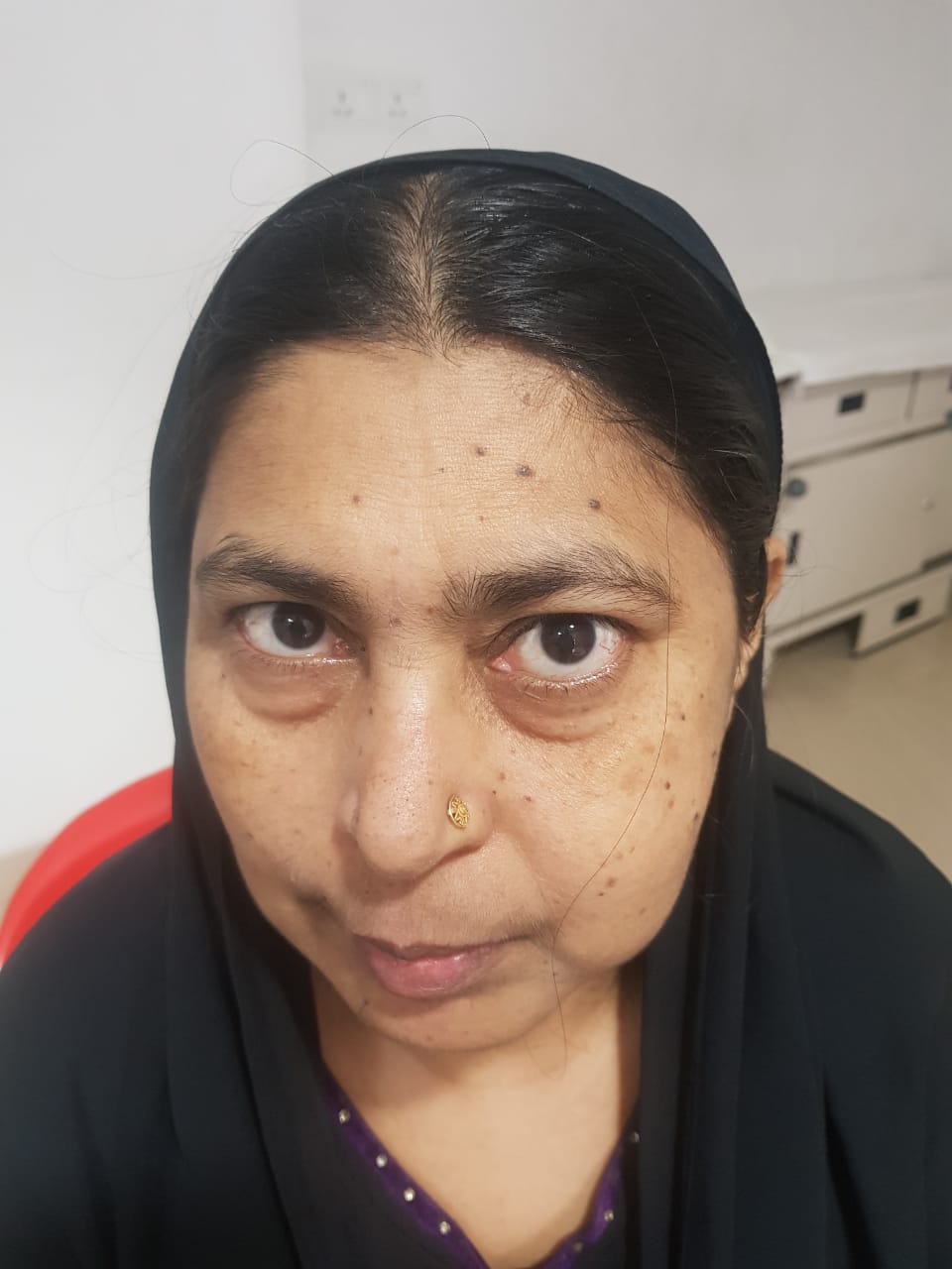
Doctors at Kohinoor Hospital, Mumbai, treated a 59-year-old woman, suffering from maxillary nerve schwannoma involving maxillary sinus. It can arise from the spinal nerves, autonomous nervous system, and the cranial nerves, except one’s optic and olfactory nerve. It may occur rarely and one may encounter facial pain, swelling of the cheek and loss of sensation over face. Dr. Sanjay Helale and Dr. Supriya Yempalle appeal for patients to immediately consult the expert if they notice any symptoms.
A 55-year-old woman, Khushnuda Khan, visited Kohinoor Hospital, Mumbai, with complaints of vague left-sided facial pain and swelling over left cheek in March 2019. She experienced the pain for almost one-and-a-half year. Initially, she represented toothache, for which she approached a dentist. Later, she was asked to undergo a dental extraction for her upper teeth, as they thought the pain was owing to dental caries (tooth decay or cavities). To add to her agony, even after opting for the dental extraction, her pain didn’t subside. Moreover, the patient continued to have dull and vague facial pain on the left side of the face. To her horror, she also noticed a swelling over the left cheek which increased over the one-and-a-half year. Though, the patient didn’t showcase any history of visual complaints like loss of vision and sensation over the left side of the face.
On conducting an examination, the doctors found a diffuse swelling over the left maxillary region of the face (the upper fixed bone of the jaw). During an intraoral examination, destruction of the upper gingivolabial sulcus (it is an area of potential space between a tooth and the surrounding gingival tissue which is lined by sulcular epithelium), on the left side was noticed. To add to her misery, there wasabnormal protrusion or displacement of an eye. While her diagnostic nasal endoscopy revealed that there was medial bowing of the lateral wall of the nose. Furthermore, her CT scan of Oromaxillary region showcased that she was suffering from Maxillary Sinusitis (the paranasal sinus is caused by a virus, bacteria or fungus). Maxillary nerve schwannoma involving maxillary sinusitis eroded the superior, inferior and posterior walls by extending laterally into the left infratemporal fossa (a complex area located at the base of the skull), Buccal fat abutting lateral pterygoid muscle (located below the zygoma and anteriorly to the ramus of the mandible surrounding the medial pterygoid and masseter muscles), and Temporalis muscle (it is located at the side of the head/skull at each of the temples). Along with this, thinning and densification of the adjoining skull base were observed.
In the MRI, it was found that there is a heterogeneous mass on the horizontal part of carotid artery (a major blood vessel in the neck that supply blood to the brain, neck, and face). Radiologically it was suspected as mucocele or cystic lesion of the maxillary sinusitis.
Dr. Sanjay Helale, Head of Department, ENT, and Head & Neck Surgery, Kohinoor Hospital says, “This wasn’t a case of diabetes/hypertension/ tuberculosis. There was no history of any medication and allergy of any drugs or any family history. But, after the routine investigations and anesthesia evaluation, the patient was taken for surgical management. The nasal cavity mass was completely removed by the combined approach that is Endonasal Endoscopic with Sublabial approach under image guidance navigation system and cranial Doppler. Diagnostic nasal endoscopy was done with 0° rigid endoscope to confirm the intranasal extension of the disease. Endoscopic medial Maxillectomy was performed to gain access to the maxillary sinusitis and help her bid adieu to it.”
He added, “Posterior wall of the maxillary sinusitis was removed throughsublabial incision to gain access to the pterygopalatine fossa (it is a neurovascular crossroad of the nasal cavity, masticator space, orbit, oral cavity, and middle cranial fossa). Profuse bleeding was observed from the maxillary artery after that. Then, the defect was packed with the medicated gauze. The gauze was removed on day 3, after treating her. There was no bleeding after the removal of gauze. The patient is fine and can do her real-world activities easily.”
";

.jpg)